Bilateral oculomotor nerve palsy secondary to bacterial meningitis
Cranial nerve palsies are a relatively uncommon complication of bacterial meningitis, occurring in approximately 4–11% of cases, and are associated with a poor prognosis when presented during hospitalization
14/03/2023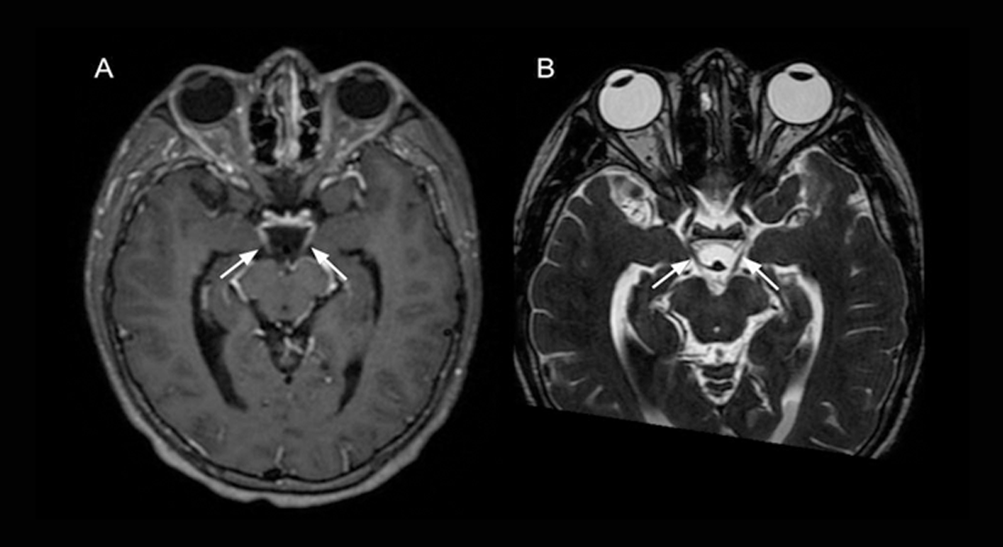
Images of the brain show bilateral enhancement and thickening of the cisternal segments of the oculomotor nerves
Kami L et al. – Bilateral nerve palsy in meningitis
Leonardo Kami[1] and Bernardo Correa de Almeida Teixeira[1]
[1]. Hospital de Clínicas da UFPR, Departamento de Radiologia e Diagnóstico por Imagem, Curitiba, PR, Brasil.
Corresponding author: Leonardo Kami. e-mail: leolkt28@gmail.com
Authors’ contribution
I would like to extend my sincere gratitude to LK for the data analysis and interpretation and for helping to draft the article, as well as to BCAT for helping to conceptualize and design the study, analyze and interpret data, and approve the final submission.
Conflict of Interest
We hereby declare that there are no conflicts of interest.
Financial Support
We have no financial support to declare. The authors provided all of the financial resources.
Orcid
Leonardo Kami: https://orcid.org/0000-0001-6111-9855
Bernardo Correa de Almeida Teixeira: https://orcid.org/0000-0003-4769-6562
A 17-year-old male was referred to a tertiary hospital with a 3-day history of headache, fever, neck stiffness, and progressive loss of consciousness. During hospitalization, he presented with bilateral mydriasis with fixed pupils, palpebral ptosis, and right horizontal gaze palsy. Cerebrospinal fluid analysis revealed xanthochromia, pleocytosis (280 WBC/microL), decreased glucose (<5.0 mg/dL), elevated protein (568.0 mg/dL), elevated lactate (16.1 mmoL/L), and latex agglutination positive for S. pneumoniae. Moreover, magnetic resonance imaging of the brain revealed intermediate signal content in the posterior segments of the lateral ventricles on FLAIR, with restricted diffusion on DWI, suggesting a dense/high protein content (Figure 1). Clinically, the most common presentation was purulent/inflammatory discharge or blood. However, in the T2* sequence, no low signals were observed, making suppurative inflammatory material secondary to ventriculitis highly likely. Leptomeningeal enhancement and thickening of the oculomotor nerves were also observed (Figure 2). After intensive care treatment with antibiotics and glucocorticoids, the patient was discharged with partial recovery from focal neurological deficits.
Cranial nerve palsies are a relatively uncommon complication of bacterial meningitis, occurring in approximately 4–11% of cases1, and are associated with a poor prognosis when presented during hospitalization2. The most common pathogen responsible is S. pneumoniae1,3. Furthermore, the most affected cranial nerves are the oculomotor, abducens, facial, and trochlear nerves, and mainly the latter, because of sensitivity to high intracranial pressure. The pathophysiological explanation is nerve compression caused by pressure on the peripheral nerve, and perineuritis caused by meningeal inflammation.
Acknowledgments
We have no acknowledgments to make.
References
- Koelman DLH, Brouwer MC, Ter Horst L, Bijlsma MW, van der Ende A, van de Beek D. Pneumococcal meningitis in adults: a prospective nationwide cohort study over a 20-year period. Clin Infect Dis. 2022;74(4):657-67.
- Weisfelt M, van de Beek D, Spanjaard L, Reitsma JB, de Gans J. Clinical features, complications, and outcome in adults with pneumococcal meningitis: a prospective case series. Lancet Neurol. 2006;5(2):123-9.
- van de Beek D, de Gans J, Spanjaard L, Weisfelt M, Reitsma JB, Vermeulen M. Clinical features and prognostic factors in adults with bacterial meningitis. N Engl J Med. 2004;351(18):1849-59. Erratum in: N Engl J Med. 2005;352(9):950.

FIGURE 1: Magnetic resonance images of the brain show intermediate signal content in the lateral ventricles at FLAIR sequence (A) with restricted diffusion at DWI (B)

FIGURE 2: Magnetic resonance images of the brain show bilateral enhancement and thickening of the cisternal segments of the oculomotor nerves at T1-weighted contrast-enhanced (A) and high-resolution T2-weighted (B) sequences. Lateral deviation of the right eyeball can be observed