Images in Infectious Diseases: A Rare Cause of Alveolar Echinococcal Metastasis
Herein, we report a rare case of alveolar echinococcosis that recurred as lung and brain metastases sometime after liver transplantation
09/02/2023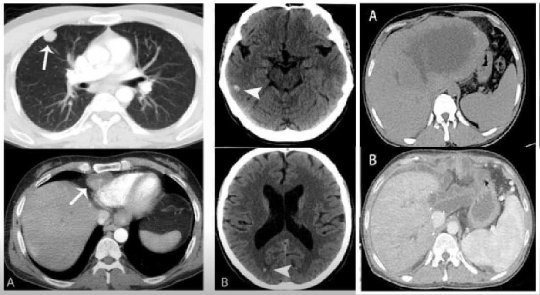
Thorax CT imaging after the transplantation (A) shows hypodense mass lesions (arrows) in the paracardiac area and within the lung parenchyma. Axial non-contrast brain CT images after the transplantation (B) show hyperdense lesions in the brain (arrowheads)
Yesilyurt M and Polat G – A Rare Cause of Alveolar Echinococcal Metastasis
Mustafa Yesilyurt[1] and Gökhan Polat[1]
[1]. Ataturk University, Medical Faculty, Department of Radiology, Erzurum, Turkey.
Corresponding author: Dr. Mustafa Yesilyurt. e-mail: drmyesilyurt@gmail.com
Authors’ contribution
MY: Conception and design of the study, Acquisition of data, Drafting the article; GP: Conception and design of the study, Final approval of the version to be submitted.
Conflict of Interest
The authors declare that they have no conflict of interest to the publication of this article.
Protection of human and animal subjects
The authors declare that no experiments were performed on humans or animals in this study.
Confidentiality of data/Right to privacy and informed consent
The authors declare that no patient data appear in this article.
Orcid
Mustafa Yesilyurt: https://orcid.org/0000-0002-7605-4940
Gökhan Polat: https://orcid.org/0000-0002-9184-8730
Received 22 November 2022 – Accepted 28 December 2022
A 27-year-old male patient was referred to the general surgery clinic of our hospital with a diagnosis of an alveolar echinococcosis cyst. After the examination and imaging, liver transplantation was performed (Figure 1). No echinococcal lesions were found in the other parts of the body before the transplant surgery. After the surgery, the patient received immunosuppressive treatment. Brain computed tomography (CT) and thoracic CT were performed 1 year after transplantation. Multiple solitary lesions were observed in the brain and lung parenchyma (Figure 2). The lesions were biopsied and diagnosed as alveolar echinococcal metastasis.
Alveolar echinococcosis is a chronic and malignant parasitic infection caused by helminth echinococcosis multilocularis1. It mostly affects the liver. Radical hepatic resection is the most effective treatment for liver cancers. Liver transplantation can be used as a curative method for the treatment of unresectable cases1. However, immunosuppressive treatments used after transplantation may rarely cause metastatic involvement in alveolar echinococcosis2. Herein, we report a rare case of alveolar echinococcosis that recurred as lung and brain metastases sometime after liver transplantation. Although resection is the potential treatment that offers the best survival in alveolar echinococcosis, it is revealed that a significant increase in metastasis and spread to surrounding tissues may occur as a result of current immunosuppressive therapy3. Therefore, the precise role and duration of perioperative immunosuppressive therapy need to be examined.
Acknowledgments
We offer our deepest thanks to the institutions that provided technical support for the
development and implementation of this study.
References
- Kern P, Bardonnet K, Renner E, Auer H, Pawlowski Z, Ammann RW, et al. European echinococcosis registry: human alveolar echinococcosis, Europe, 1982–2000. Emerg Infect Dis. 2003;9(3):343-9. Available from: https://doi.org/10.3201/eid0903.020341
- Caire Nail L, Rodríguez Reimundes E, Weibel Galluzzo C, Lebowitz D, Ibrahim YL, Lobrinus JA, et al. Disseminated alveolar echinococcosis resembling metastatic malignancy: a case report. 2017;11(1):113. Available from: https://doi.org/10.1186/s13256-017-1279-2
- Lachenmayer A, Gebbers D, Gottstein B, Candinas D, Beldi G. Elevated incidence of alveolar echinococcosis in immunocompromised patients. Food Waterborne Parasitol. 2019;16:e00060. Available from: https://doi.org/10.1016/j.fawpar.2019.e00060
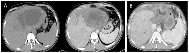
FIGURE 1: Axial non-contrast and contrast-enhanced computed tomography (CT) images (A) show a hypodense mass lesion in the left lobe of the liver before transplantation. Axial contrast-enhanced CT imaging after liver transplantation (B)

FIGURE 2: Thorax CT imaging after the transplantation (A) shows hypodense mass lesions (arrows) in the paracardiac area and within the lung parenchyma. Axial non-contrast brain CT images after the transplantation (B) show hyperdense lesions in the brain (arrowheads)