IMAGES IN INFECTIOUS DISEASES: Oral presentation of histoplasmosis in non-HIV immunocompromised patient after cardiac transplant: First Brazilian case report
Histoplasmosis is acquired mainly by the inhalation of fungi and occurs in both immunocompromised and immunocompetent individuals. Human-to-human transmission is rare and is reported only in the context of organ transplantation
09/01/2023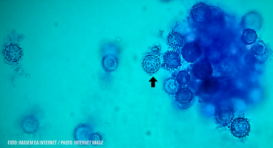
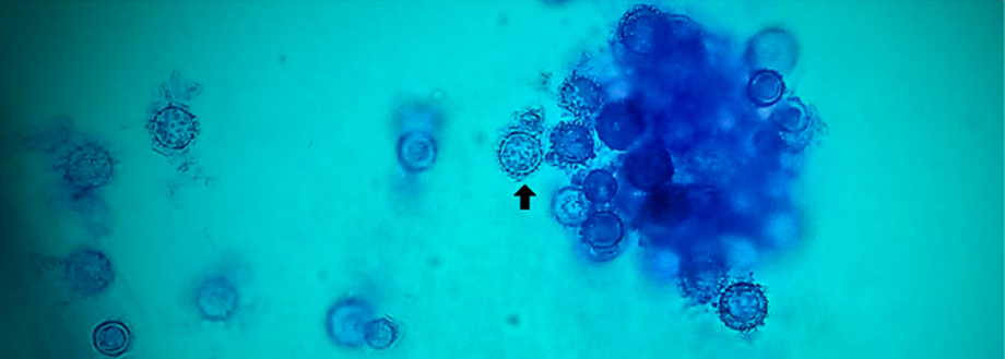
Fungal structures of the isolate were obtained from microculture on cornmeal agar at room temperature. Micromorphology shows many macroconidia (black arrow). Observation by optical microscopy at 400x magnification using cotton blue stain
Araújo MRB et al. – Disseminated histoplasmosis in cardiac transplanted patient
Max Roberto Batista de Araújo[1], Louisy Sanches dos Santos[2] and Lincoln de Oliveira Sant’Anna[2]
[1]. Instituto Hermes Pardini, Núcleo Técnico Operacional, Microbiologia, Vespasiano, MG, Brasil.
[2]. Universidade do Estado do Rio de Janeiro, Faculdade de Ciências Médicas, Departamento de Microbiologia, Imunologia e Parasitologia, Rio de Janeiro, RJ, Brasil.
Corresponding author: Max Roberto Batista de Araújo. e-mail: max_barau@hotmail.com
Authors’ contribution
LOS: Conceptualization, Data curation, Formal analysis, Writing-original draft, and Writing-review & editing; LSS: Conceptualization, Data curation, Formal analysis, and Writing-original draft; MRBA: Conceptualization, Data curation, Formal analysis, Investigation, Methodology, Project administration, and Writing-review & editing.
Compliance with ethical standards
Conflict of interest
The authors declare that they have no conflict of interest.
Ethical approval
Since the study was a retrospective analysis of laboratory data collected and no additional investigations were performed with submitted specimen, no ethical approval was applied.
Financial Support
Funding information is not applicable. No funding was received.
Orcid
Max Roberto Batista de Araújo: https://orcid.org/0000-0002-3293-8496
Louisy Sanches dos Santos: https://orcid.org/0000-0002-5303-6395
Lincoln Oliveira Sant’Anna: https://orcid.org/0000-0002-6933-8961
Received 29 September 2022 – Accepted 22 November 2022
A 56-year-old Brazilian man with systemic arterial hypertension and type 2 diabetes mellitus was affected by ischemic heart disease that led to heart transplantation. He underwent angioplasty with stent placement and was taking immunosuppressants. Four months later, he was readmitted to the intensive care unit with an oral ulcer, fever, and holocranial headache. The biopsies of the oral lesions (Figure 1) and transbronchial showed granulomatous inflammation (Figure 2).
Laboratory tests yielded negative results for HIV, hepatitis virus, tuberculosis, toxoplasmosis, Cytomegalovirus, Cryptococcus spp. and Paracoccidioides spp. Blood and oral fragment samples were collected simultaneously with the application of amphotericin B lipid complex for fungal cultures and showed growth after approximately 27 days. The microculture of white colonies suggested Histoplasma spp. (Figure 3), a diagnosis confirmed by immunodiffusion tests and mass spectrometry by MALDI-TOF. This treatment was then switched to itraconazole. The patient showed clinical improvement and was discharged after 37 days of hospitalization.
Histoplasmosis is acquired mainly by the inhalation of fungi and occurs in both immunocompromised and immunocompetent individuals. Human-to-human transmission is rare and is reported only in the context of organ transplantation. Once the fungus has invaded the host, it evades immune defenses, finds a niche for its growth and reproduction, and may spread or develop a latent state within granulomas1. Oral manifestations in the disseminated form are particularly associated with HIV infection2,3. In conclusion, oral involvement is extremely rare in immunocompetent or non-HIV-infected patients, confirming that the case reported here is rare.
References
- Mittal J, Ponce MG, Gendlina I, Nosanchuk JD. Histoplasma Capsulatum: Mechanisms for Pathogenesis. Curr Top Microbiol Immunol. 2019;422:157-91. Available from: https://doi.org/10.1007/82_2018_114
- Ferrisse TM, Rocha AFL, Miotto LN, Lança MLA, Massucato EMS, Bufalino A. Disseminated Histoplasmosis Infection in HIV-Negative Patients: Series Case and Literature Review. Curr Fungal Infect Rep. 2021;15:17-22. Available from: https://doi.org/10.1007/s12281-021-00414-w
- Kamboj M, Rathee R, Narwal A, Devi A, Gupta A, Sivakumar N. Primary Oral Histoplasmosis in Immunocompetent Host: Case Series with Review of Literature. Indian J Otolaryngol Head Neck Surg. 2021. Available from: https://doi.org/10.1007/s12070-021-02974-2
FIGURE 1: Biopsies of oral lesions and transbronchial showing granulomatous inflammation due to fungal disease. (a) Panoramic image of ulcerated mucosa infiltrated by a mixed inflammatory exudate (original magnification 1x–scanner: Aperio®). (b) Panoramic image with a focus on the ulceration (original magnification 4x–scanner: Aperio®). (c) Detail of the ulcer permeated by a mixed inflammatory infiltrates (original magnification 10x–scanner: Aperio®). (d) Clear cytoplasmic cells (black arrow) with small fungal structures, mainly in the subepithelial area (10x magnification–scanner: Aperio®).
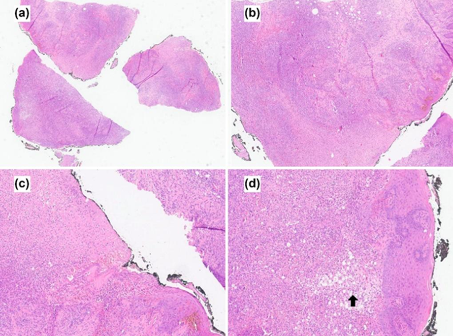
FIGURE 2: Special Gomori-Grocott staining. (a) Fungal structures (black arrow) in the cytoplasm of clear cytoplasmic cells (20x magnification–scanner: Aperio®). (b) Fungal structures (black arrow) within the cytoplasm of clear cytoplasmic cells (original magnification 40x – Scanner: Aperio®)
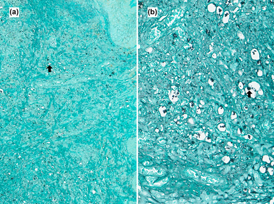
FIGURE 3: Fungal structures of the isolate were obtained from microculture on cornmeal agar at room temperature. Micromorphology shows many macroconidia (black arrow). Observation by optical microscopy at 400x magnification using cotton blue stain